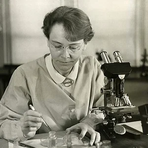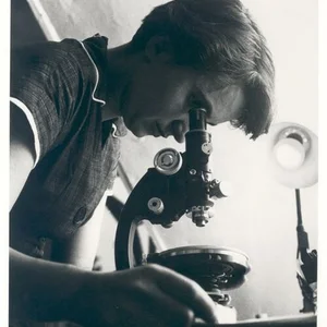

British molecular biologist (1920–1958), Rosalind Franklin made essential contributions to our understanding of DNA structure through her X-ray crystallography work. She is best known for Photo 51, a landmark image that revealed the double helix structure of DNA.
Frequently asked questions
Key Facts
- 1941–1947: Advanced studies in physical chemistry and research in coal crystallography
- 1951–1952: Production of the famous Photo 51, showing the structure of DNA by X-ray diffraction at King's College London
- 1953: Her critical data contributed to Watson, Crick, and Wilkins's elucidation of the double helix structure of DNA
- 1955: Pioneering work on the structure of viruses and RNA
- 1958: Death at age 37 from cancer, before official recognition of her foundational role
Works & Achievements
An X-ray diffraction image revealing the double helix structure of DNA. This crucial photograph provided visual proof of DNA's molecular geometry and was instrumental in the model proposed by Watson, Crick, and Wilkins.
A series of experiments using X-ray crystallography to analyze the structure of deoxyribonucleic acid. This work revealed the dimensions and configuration of the DNA molecule with unprecedented precision.
Crystallographic studies of the structure of the tobacco mosaic virus. Franklin determined that this virus had a helical structure, contributing to the understanding of viral architecture.
Franklin published several major scientific papers detailing her discoveries on the structures of DNA and proteins. These publications laid the scientific foundations for future generations of molecular biologists.
Franklin refined and applied X-ray crystallography methods to the study of biological molecules. Her technical innovations enabled a resolution of complex molecular structures that had never before been achieved.
Anecdotes
Rosalind Franklin captured the famous 'Photo 51' in 1952, an X-ray diffraction image that clearly revealed the double helix structure of DNA. The photograph was so sharp and detailed that Watson and Crick used it as key evidence for their DNA model, even though Franklin was not initially credited for this decisive contribution.
At the age of 31, Rosalind Franklin was diagnosed with cancer in 1956, most likely caused by her prolonged exposure to X-rays during her scientific experiments. She continued working on her research despite her illness and died on April 16, 1958 — four years before Watson, Crick, and Wilkins received the Nobel Prize for their work on DNA.
Franklin was known as an independent woman with a deep passion for research: she was fluent in several languages (French, German, and Hebrew) and had to leave France in 1940 to escape the Nazi occupation before continuing her scientific studies in Britain. Her determined yet sometimes reserved personality earned her a reputation as a scientist who held herself and her colleagues to exacting standards.
Although her crucial role in discovering the structure of DNA was long overlooked, Rosalind Franklin also conducted pioneering research into the structure of viruses and RNA. Her early work on the chemistry of coal contributed to the British war effort during the Second World War.
The laboratory where Franklin carried out her DNA experiments was located at King's College London, a prestigious scientific environment where she nonetheless had to contend with prejudice as a woman in science. Despite these obstacles, her experimental data was so precise and rigorous that it revolutionized molecular biology for generations to come.
Primary Sources
The X-ray diffraction patterns of sodium thymonucleate fibres have been studied quantitatively, and the results suggest a possible structure for this important protein complex. The very regular structure and the uniformity of the pattern suggest a regular, ordered arrangement of the atoms in the fibre.
Evidence has been presented for a helical structure of the deoxyribonucleic acid molecule based on X-ray diffraction data. The angle of the helix and the number of residues per turn have been determined from the observed diffraction patterns.
I have obtained very good X-ray diffraction photographs showing a highly regular structure. The crystallographic data strongly suggest a helical configuration of the DNA molecule.
The structure of deoxyribonucleic acid displays remarkable characteristics that can only be explained by a regular, ordered arrangement of its constituent molecules.
Key Places
Rosalind Franklin's birthplace in 1920. She grew up in a prosperous Jewish family and received an excellent education before going on to pursue her university studies.
The institution where Rosalind Franklin conducted her X-ray crystallography research between 1951 and 1953. It was here that she produced the famous Photo 51, a crucial piece of evidence for the double helix structure of DNA.
The institution where Franklin worked from 1953 to 1958 on viruses and nucleic acids. There she continued her groundbreaking crystallography research with greater scientific freedom.
Where Rosalind Franklin completed a formative fellowship in 1947–1948 at the Laboratoire Central de Chimie Physique. She refined her X-ray crystallography techniques during her time there.
The prestigious institution where Franklin's discoveries about DNA were recognised and built upon. Watson, Crick, and Wilkins — who went on to receive the Nobel Prize — worked nearby at Cambridge.
Liens externes & ressources
Références
Œuvres
Photographie 51 (Photo 51)
Mai 1952
Article 'Molecular Configuration in Sodium Thymonucleate', Nature
25 avril 1953
Travaux sur la structure du virus de la mosaïque du tabac (TMV)
1955-1958
Thèse de doctorat sur la structure des charbons
1945
Travaux sur la structure du virus de la mosaïque du turnip (TYMV)
1956-1958